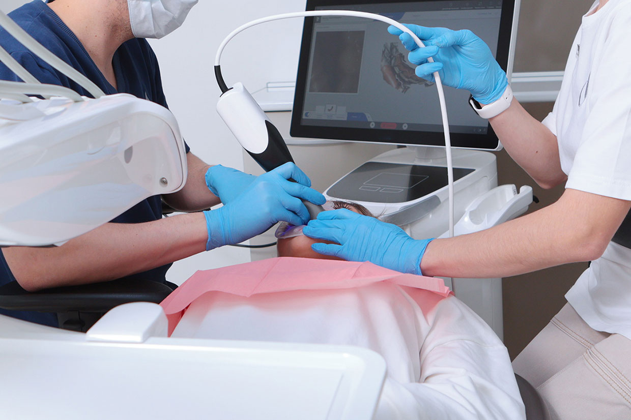
What if your dental team could gather additional information about a developing cavity before it becomes a larger problem? Modern diagnostic technology is giving dentists more ways to examine teeth beyond what can be seen during a visual examination alone. Near Infrared Imaging, commonly called NIRI, uses near infrared light to create detailed images that may help identify early signs of tooth decay. Patients interested in this technology can learn more about the practice’s advanced dental technology and how digital tools can support preventive care.
NIRI cavity detection in McHenry is especially useful because early decay does not always hurt. A tooth can look and feel completely normal while changes are developing beneath or between visible surfaces. By combining careful examination with modern imaging, dentists can gather more information before deciding whether a tooth simply needs monitoring, stronger preventive care, or restorative treatment.
What Is NIRI Cavity Detection?
NIRI stands for Near Infrared Imaging. The technology uses near infrared light to create images of dental structures without relying solely on visible light.
Different parts of a tooth interact with near infrared light in different ways. Healthy enamel and areas affected by decay may therefore appear differently on the resulting image. This can give the dentist another way to examine areas where early cavities may be difficult to identify through visual inspection alone.
NIRI cavity detection is noninvasive. It does not involve drilling, injections, or removing tooth structure. Instead, the imaging process gathers diagnostic information that can be considered alongside the rest of the dental examination.
It is important to understand that NIRI does not automatically diagnose every cavity by itself. Dental diagnosis still depends on professional interpretation, examination findings, symptoms, dental history, and other imaging when appropriate.
Why Early Cavity Detection Matters
Tooth decay is a progressive process. It begins when acids produced by bacteria contribute to mineral loss from the tooth surface. At first, the changes may be limited to enamel and may not cause discomfort.
As decay progresses, it can move deeper into the tooth. Once it reaches softer dentin, the cavity may expand more quickly. Eventually, untreated decay may approach the nerve-containing pulp and contribute to pain, infection, or the need for more extensive treatment.
This is why early detection matters. Finding suspicious areas sooner can give patients and dentists more options.
In some early situations, a dentist may recommend preventive measures and close monitoring. If a true cavity has progressed to the point where restoration is needed, treatment may involve a relatively conservative dental filling.
Waiting until a tooth hurts can allow decay to become significantly larger before treatment begins.
How Does Near Infrared Imaging Work?
NIRI uses light in a wavelength that is outside the range people normally see. The technology directs this light through dental structures and captures the resulting image.
Because sound tooth structure and areas of demineralization can transmit or scatter the light differently, the image may reveal changes that deserve additional attention.
The process can be particularly useful when examining certain surfaces between teeth. These contact areas can be difficult to see directly because neighboring teeth block the view.
During a dental visit, the dentist reviews the NIRI images in the context of the complete examination. If an area appears suspicious, additional evaluation may be recommended before deciding on treatment.
Therefore, NIRI cavity detection works best as one component of a comprehensive diagnostic approach.
Does NIRI Replace Dental X-Rays?
No. NIRI and dental X-rays provide different kinds of information.
Near infrared imaging is useful for examining certain dental structures and detecting changes associated with tooth decay. Dental X-rays, meanwhile, can show roots, bone, areas around restorations, and other structures that NIRI cannot evaluate in the same way.
For example, a dentist investigating bone levels around teeth or an infection near a root may need radiographic information rather than near infrared imaging.
Likewise, NIRI may provide another useful perspective when evaluating areas between teeth for early decay.
The technologies can complement each other. The dentist chooses which diagnostic tools are appropriate according to the patient’s history, symptoms, cavity risk, and treatment needs.
Can NIRI Find Cavities Between Teeth?
One of the challenges of cavity detection is evaluating the contact areas where neighboring teeth touch. These spaces are difficult to inspect directly with the eyes.
NIRI may help provide information about changes in these areas because near infrared light can be used to create an image of tooth structure that is otherwise difficult to visualize.
However, the dentist must still interpret what the image means. Not every dark or unusual area automatically represents decay requiring a filling.
The location, depth, appearance, previous records, and overall cavity risk all influence treatment decisions.
This careful approach is especially important because modern preventive dentistry aims to preserve as much healthy tooth structure as possible.
Benefits of NIRI Cavity Detection
NIRI cavity detection can provide several advantages when it is used appropriately as part of a dental examination.
- Earlier identification of suspicious areas: NIRI may help reveal changes before they become obvious to the patient.
- Noninvasive imaging: The technology gathers information without drilling or altering the tooth.
- Comfortable examination: Near infrared imaging is designed to collect diagnostic information without causing tooth pain.
- Helpful views between teeth: NIRI may provide additional information about contact areas that are difficult to inspect visually.
- Support for preventive decisions: Earlier information can help the dentist determine whether an area should be monitored, protected, or treated.
- Visual patient education: Digital images can make it easier for patients to understand areas the dentist is monitoring.
- Complementary diagnostic information: NIRI can be used alongside visual examinations, dental X-rays, and other diagnostic methods.
- Focus on tooth preservation: Detecting decay earlier supports the goal of managing problems before larger amounts of tooth structure are affected.
The real value of NIRI cavity detection is not simply that it is newer technology. Its value comes from giving the dentist another source of information when evaluating the health of a tooth.
Who May Benefit From NIRI Cavity Detection?
NIRI may be useful for many patients, particularly when the dental team wants additional information about possible early decay.
Patients with a history of frequent cavities may benefit from closer monitoring. Likewise, people with crowded teeth or tight contacts may have areas that are especially difficult to inspect visually.
Patients with dry mouth may also face increased cavity risk because saliva plays an important protective role in the mouth.
Orthodontic treatment, dietary habits, inconsistent oral hygiene, and previous restorative work can also influence cavity risk.
However, a patient does not need to be considered high risk for NIRI to be useful. The dentist determines when the technology adds helpful information to the examination.
What Happens if NIRI Shows a Suspicious Area?
A suspicious image does not necessarily mean the tooth will be treated immediately.
The dentist first considers what the image shows in combination with the physical examination. Additional dental images may be reviewed when appropriate.
If the change appears very early and the tooth surface remains intact, preventive strategies may sometimes be recommended. These could include improved brushing, fluoride exposure, changes in snacking patterns, and more careful monitoring.
If a cavity has progressed and tooth structure has been damaged, a filling may be recommended to remove decay and restore the area.
The goal is to match treatment to the actual stage of the problem rather than automatically treating every suspicious image the same way.
Can Early Tooth Decay Be Reversed?
The earliest stage of enamel demineralization may sometimes be managed before a physical cavity forms. Enamel can gain minerals from saliva and fluoride under favorable conditions.
However, once decay has created a structural hole in the tooth, brushing and fluoride cannot simply rebuild the missing enamel.
This distinction is one reason early detection can be valuable. Identifying demineralization before substantial structural damage occurs may create an opportunity for more preventive management.
The dentist can help determine whether an area represents early mineral loss, an established cavity, staining, or another finding.
How Does NIRI Fit Into a Routine Dental Examination?
A comprehensive dental examination may involve several diagnostic methods. The dentist visually examines teeth and gums, checks existing restorations, evaluates areas of concern, and considers symptoms reported by the patient.
When additional information is needed, imaging may be incorporated into the visit.
NIRI can add another perspective when evaluating tooth structure for possible decay. Meanwhile, conventional dental X-rays may be used to examine roots, bone, and other areas.
This layered approach reduces reliance on any single diagnostic method.
Patients can support this process by maintaining routine dental cleanings and examinations, even when their teeth feel healthy.
What Can You Do to Reduce Your Cavity Risk?
Technology can help detect tooth decay, but daily habits remain essential for prevention.
Brush twice each day with fluoride toothpaste and clean between teeth daily. Interdental cleaning is particularly important because cavities can develop in areas that toothbrush bristles do not reach effectively.
Try to reduce frequent exposure to sugary beverages and snacks. Frequency matters because repeated carbohydrate exposure gives oral bacteria more opportunities to produce acids.
Drinking water throughout the day also supports saliva and helps rinse loose debris from the mouth.
Finally, maintain the preventive visit schedule recommended for your individual risk level. Regular monitoring can make it easier to identify changes before symptoms develop.
Frequently Asked Questions About NIRI Cavity Detection
Does NIRI cavity detection hurt?
No. Near infrared imaging is noninvasive and is designed to gather diagnostic information without drilling, injections, or removing tooth structure.
Does NIRI use the same technology as a dental X-ray?
No. NIRI uses near infrared light, while dental X-rays use radiographic imaging. The two methods provide different types of diagnostic information.
Can NIRI replace all dental X-rays?
No. Dental X-rays can show roots, bone, and other structures that near infrared imaging cannot evaluate in the same way. Dentists select imaging based on the information needed.
Can NIRI detect cavities before they hurt?
It may help identify suspicious changes associated with tooth decay before the patient notices pain. However, the dentist must interpret the images together with other examination findings.
If NIRI shows decay, will I automatically need a filling?
Not necessarily. Treatment depends on whether the area represents early demineralization, an established cavity, or another finding. The dentist determines the appropriate approach after a complete evaluation.
Detect Dental Changes Earlier in McHenry
NIRI cavity detection gives dentists another way to examine tooth structure for early signs of decay. By combining near infrared imaging with visual examination, dental X-rays when appropriate, and individualized cavity risk assessment, the dental team can make more informed decisions about prevention, monitoring, and treatment.
To learn more about NIRI cavity detection in McHenry, call Unique Smiles of McHenry at (815) 385-0777. Visit our office at 4101 W Shamrock Ln, McHenry, IL 60050, or Book an Appointment for personalized preventive dental care.