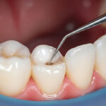
Have you ever heard a dentist describe a tiny crack, worn filling, or irritated area and wished you could see exactly what they were talking about? An intraoral camera can make that conversation much easier. This small handheld device captures close-up images from inside the mouth, allowing patients and dental professionals to view teeth and gums in greater detail. Intraoral cameras are one of several types of dental technology that can support diagnosis, patient education, and treatment planning.
Intraoral cameras in McHenry can be particularly helpful because many dental concerns occur in places that are difficult to view in a bathroom mirror. Back teeth, the tongue side of the lower teeth, restoration margins, and small areas near the gumline may all be easier to understand when enlarged on a screen. Instead of relying only on a verbal description, patients can become more involved in conversations about their oral health.
What Is an Intraoral Camera?
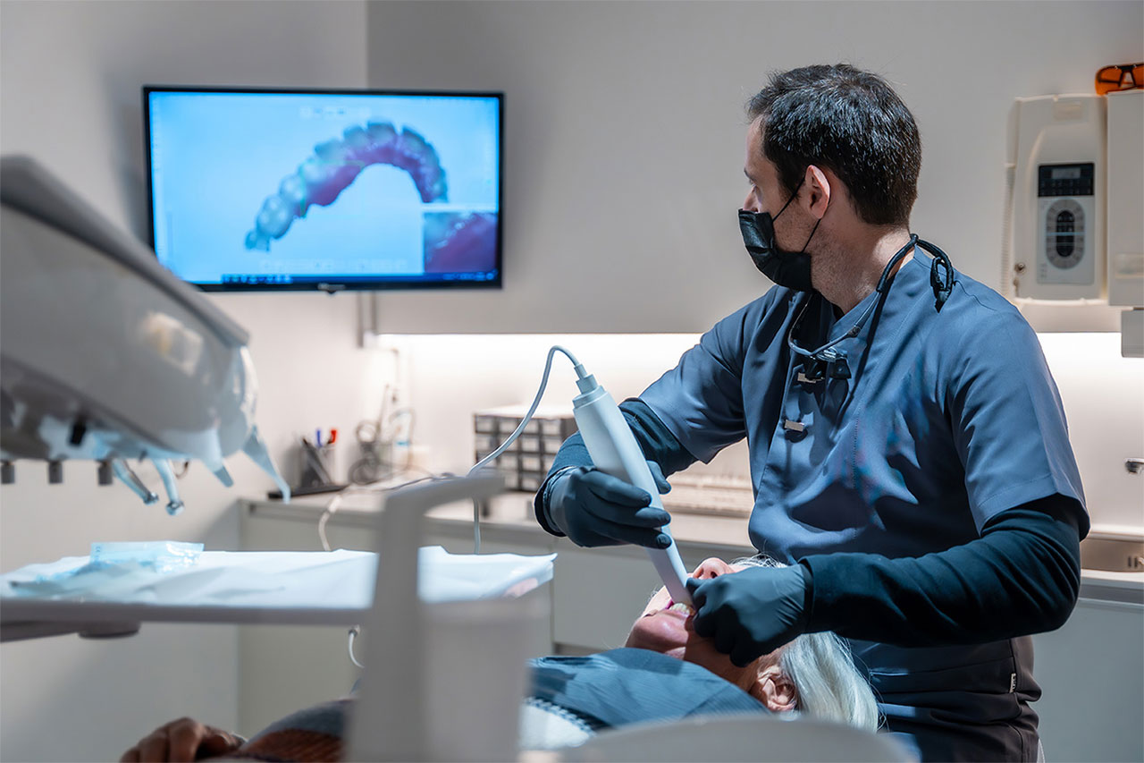
An intraoral camera is a compact digital camera designed to capture images inside the mouth.
The device is often similar in size to a pen. Its small design allows the dental professional to position it near different teeth, gums, and oral structures.
Images can then be displayed on a monitor. Depending on the dental system, they may also be stored as part of the patient’s records for future comparison.
Unlike a traditional dental mirror, an intraoral camera can enlarge what is being viewed. This makes tiny details easier to see and explain.
The camera does not diagnose a dental condition by itself. Instead, it provides visual information that the dentist combines with the clinical examination, dental imaging, symptoms, and history.
What Can an Intraoral Camera Show?
An intraoral camera can capture visible structures throughout the mouth.
Depending on the area being examined, it may help document:
- Visible cavities or suspicious areas
- Cracked or chipped teeth
- Worn tooth surfaces
- Old or damaged fillings
- Areas of gum inflammation
- Plaque or tartar accumulation
- Recession around the gumline
- Discoloration
- Changes around crowns or bridges
- Other visible oral findings
The ability to enlarge these areas can make subtle changes easier to discuss.
For example, a patient may understand why a filling needs monitoring when they can see a worn margin on a large screen rather than simply being told that the restoration looks old.
How Is an Intraoral Camera Different From an Intraoral Scanner?
The names sound similar, but the technologies serve different purposes.
An intraoral camera captures photographs or video-like images of visible structures in the mouth. Its primary value is documentation, visualization, and patient education.
An intraoral scanner captures detailed three-dimensional surface information. Those digital models can be used for treatments such as crowns, bridges, aligners, and other restorations.
Therefore, one technology does not simply replace the other.
An intraoral camera helps the dentist and patient closely view a particular tooth or tissue. An intraoral scanner creates a broader digital model of the shape and position of teeth.
Depending on the treatment, both technologies may provide useful information.
Why Is Seeing the Problem Helpful?
Dental terminology can be difficult to visualize. Words such as fracture line, recurrent decay, gum recession, worn restoration, or calculus may not mean much to someone who has never seen those conditions.
A close-up image can make the explanation more concrete.
For example, if the dentist sees a crack near an old filling, the image can be displayed while the treatment options are discussed. If gum inflammation is present, patients may be able to see redness or buildup that needs more attention.
This does not mean patients must diagnose themselves. Instead, images can make professional explanations easier to understand.
Better understanding can also lead to more useful questions and more confident treatment decisions.
How Can Intraoral Cameras Help During Preventive Visits?
Preventive dentistry focuses on maintaining health and identifying changes before they become larger problems.
During dental cleanings and examinations, an intraoral camera may help document visible areas that deserve monitoring.
For instance, the dental team may photograph a small enamel crack, a worn restoration, recession near a tooth, or an area where plaque repeatedly accumulates.
At a future visit, another image may help show whether the condition has changed.
This type of documentation can provide useful context because memory alone is not always precise enough to determine whether an area looks exactly the same months later.
Can an Intraoral Camera Detect Cavities?
An intraoral camera can help a dentist see certain visible signs associated with decay, such as discoloration, surface breakdown, or an obvious cavity.
However, it cannot reveal every cavity.
Decay can develop between teeth or beneath surfaces that the camera cannot directly see. Dental X-rays, NIRI, or other diagnostic tools may therefore be needed.
The camera should be thought of as one part of the examination rather than a replacement for other imaging.
When multiple diagnostic methods are combined, the dentist can gather a more complete picture of the tooth.
Benefits of Intraoral Cameras
Intraoral cameras can make dental visits more visual, collaborative, and easier to understand.
- Close-up visualization: Small details can be enlarged on a monitor for easier viewing.
- Better patient education: Patients can see the area being discussed instead of relying only on a verbal explanation.
- Useful documentation: Images can become part of the dental record and may help monitor visible changes over time.
- Support for treatment planning: Photographs can provide additional information about tooth surfaces, restorations, and gum tissues.
- Improved communication: Dentist and patient can look at the same image while discussing concerns and options.
- Comfortable imaging: The device is small and designed for use inside the mouth.
- Immediate viewing: Images can often be displayed shortly after they are captured.
- Motivation for home care: Seeing plaque, tartar, or gum inflammation can help patients understand which areas need more attention.
The main benefit of an intraoral camera is clarity. Patients often feel more comfortable making decisions when they understand what the dentist is seeing.
Does an Intraoral Camera Hurt?
An intraoral camera is noninvasive and does not cut, drill, or alter the teeth.
The device is positioned around different areas of the mouth while images are captured.
Patients with a sensitive gag reflex should let the dental team know. The camera is small, but back areas of the mouth can still feel sensitive for some people.
The dental professional can often adjust positioning or take brief pauses when necessary.
For most patients, capturing intraoral photographs is a straightforward part of the dental examination.
Can Intraoral Images Help Monitor Dental Work?
Yes. Fillings, crowns, bridges, and other restorations change over time just as natural teeth do.
A dentist may use an intraoral camera to document visible restoration margins, surface wear, cracks, chips, or changes in nearby gum tissue.
Future images can then provide a point of comparison.
This does not mean every cosmetic change requires treatment. A restoration may show minor wear while continuing to function well.
However, monitoring allows the dentist to decide whether an area remains stable or whether repair or replacement should be discussed.
How Do Intraoral Cameras Support Gum Health?
Gum problems often begin in areas patients cannot easily see.
An intraoral camera can help display redness, swelling, recession, plaque, or tartar around visible portions of the teeth.
This can be useful when explaining why a particular area bleeds during brushing or why a patient needs to improve cleaning near a restoration.
However, photographs do not measure periodontal pockets or show all supporting bone changes.
A complete gum evaluation may also involve periodontal measurements, dental X-rays, and professional examination.
Again, the camera adds visual information rather than replacing the rest of periodontal diagnosis.
Can Intraoral Cameras Help With Cosmetic Dentistry?
Dental photographs are useful when evaluating the visible appearance of teeth and gums.
Before cosmetic treatment, images can help document tooth shape, color, alignment, wear, spacing, and other characteristics.
They may also help patients communicate what they would like to change.
For example, someone considering bonding may be concerned about a small chip, while another patient may want to discuss discoloration or uneven tooth edges.
Close-up photographs can make those conversations more specific.
However, cosmetic recommendations should still be based on oral health, bite relationships, and realistic treatment goals.
Can You Keep or View Your Intraoral Images?
Images captured during a dental visit may be displayed during the appointment so the dentist can explain findings.
How the records are stored or shared depends on the dental office’s systems and policies.
The important point is that patients can benefit from seeing the images during the discussion.
If you do not understand what you are looking at, ask the dentist to point out the specific area of concern and explain what a healthy tooth or restoration would look like by comparison.
Visual information is most useful when it is paired with clear professional explanation.
Does Seeing an Image Mean Treatment Is Definitely Needed?
No. A photograph documents appearance, but the dentist still needs to determine the clinical significance of what is visible.
A small crack may be superficial and require monitoring. A stain may not be a cavity. An older filling may remain functional even if its surface no longer looks new.
Likewise, some serious dental conditions cannot be diagnosed from an intraoral photograph alone.
Treatment recommendations should come from the complete examination rather than the image by itself.
Patients should feel free to ask why treatment is recommended, what the image shows, and whether monitoring is a reasonable alternative.
How Intraoral Cameras Encourage Collaborative Dental Care
Modern dentistry increasingly emphasizes patient involvement in treatment decisions.
An intraoral camera supports that approach by allowing patients to see many of the same visible details their dentist sees.
Instead of hearing that a tooth has a problem somewhere near the back of the mouth, the patient can often view the specific area on a monitor.
This can make conversations about urgency, treatment alternatives, and home care more meaningful.
Patients do not need dental training to participate in their care. They simply need clear information and the opportunity to ask questions.
Frequently Asked Questions About Intraoral Cameras
Is an intraoral camera an X-ray?
No. An intraoral camera captures visible images of teeth and tissues. Dental X-rays provide information about structures beneath or between visible surfaces.
Does an intraoral camera hurt?
No. The camera is a small, noninvasive device designed to capture images inside the mouth without cutting or drilling.
Can an intraoral camera find every cavity?
No. It can show visible changes, but cavities between teeth or beneath surfaces may require dental X-rays, NIRI, or other diagnostic methods.
Why does my dentist take pictures of old fillings?
Photographs can help document the condition of restorations and provide a reference for comparing wear, cracks, margins, or other visible changes over time.
Can I see the pictures during my appointment?
Intraoral images can often be displayed on a monitor so the dentist can explain findings and treatment considerations more clearly.
See Your Oral Health More Clearly in McHenry
Intraoral cameras turn dental conversations into something patients can see. By providing enlarged views of teeth, gums, restorations, and other visible structures, these cameras can support diagnosis, monitoring, education, and treatment planning. Most importantly, they can help patients better understand why a dentist is recommending a particular type of care.
To experience technology-focused dental care in McHenry, call Unique Smiles of McHenry at (815) 385-0777. Visit us at 4101 W Shamrock Ln, McHenry, IL 60050, or Book an Appointment for your next dental examination.