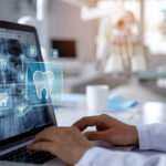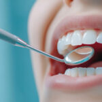

Do you remember dental impressions that involved sitting still with a tray of impression material in your mouth? Digital dentistry has created another way to capture detailed information about teeth and bite relationships. With intraoral scanning, a small handheld scanner can create a three-dimensional digital model of the mouth that helps dentists plan many types of restorative and cosmetic care.
Intraoral scanning in McHenry can make the process of collecting dental records more efficient and comfortable for many patients. Instead of relying only on traditional impression materials, the scanner captures a series of detailed digital images and combines them into a virtual model. The resulting scan can help with the design of crowns, bridges, implant restorations, and other treatments that depend on precise information about tooth shape and position.
What Is Intraoral Scanning?
Intraoral scanning is a digital method of recording the surfaces of teeth and surrounding structures. A compact wand is moved carefully around the mouth while the system captures detailed images.
Software then combines the images to create a three-dimensional representation that can be viewed on a computer screen. The dental team can rotate, enlarge, and examine the model from multiple angles.
Because intraoral scanning produces digital information, the records can often be transferred electronically when a dental laboratory or another part of the treatment process needs them.
However, intraoral scanning does not replace every diagnostic tool. Dental X-rays, visual examination, periodontal measurements, and other records each provide different information. Instead, intraoral scanning adds another useful perspective when the shape and position of teeth must be recorded accurately.
How Does an Intraoral Scanner Work?
The process usually begins with the teeth being prepared for scanning. Depending on the treatment, the dental team may dry certain surfaces or gently move soft tissues out of the way so the scanner has a clear view.
The small scanner wand is then guided around the teeth. As it moves, the system captures digital information from different angles. The patient typically keeps their mouth open while the dental professional scans the necessary areas.
The software assembles those images into a digital model. If an area is incomplete or unclear, the dental team can often rescan that specific portion instead of starting the entire process again.
Once the intraoral scanning is complete, the digital model can be reviewed immediately. This provides an opportunity to verify the information before the appointment moves forward.
What Dental Treatments Can Use Intraoral Scanning?
Intraoral scanning can be useful for several types of dental treatment. The exact applications depend on the technology, clinical situation, and treatment plan.
Common uses may include:
- Dental crowns
- Dental bridges
- Selected dental implant restorations
- Orthodontic planning
- Clear aligner treatment
- Night guards and other oral appliances
- Monitoring tooth position or wear
- Creating records of the bite and tooth surfaces
For example, when a patient needs a dental bridge, accurate information about the prepared teeth, surrounding teeth, and bite is essential. Intraoral scanning can help provide that information to support the design process.
Similarly, digital records can be valuable when planning restorations supported by dental implants. The dentist determines which digital and imaging tools are appropriate for each stage of care.
How Is Intraoral Scanning Different From Traditional Dental Impressions?
Traditional impressions typically use trays filled with a soft material that records the shape of the teeth as it sets. Traditional methods have been used successfully for many years and may still be appropriate in some situations.
Intraoral scanning takes a different approach. Instead of creating an immediate physical mold, the scanner produces a digital model.
For many patients, avoiding a tray full of impression material can make the experience more comfortable. This can be especially helpful for people with a sensitive gag reflex or those who dislike the feeling of impression material in the mouth.
Digital scanning can also make it easier to correct a small incomplete area. If part of a conventional impression is unusable, a new impression may sometimes be necessary. With intraoral scanning, the dental professional may be able to rescan only the area that needs more information.
Neither method should be viewed as automatically right for every clinical situation. The dentist selects the approach that provides suitable information for the planned treatment.
Benefits of Intraoral Scanning
Intraoral scanning offers several advantages for patients and dental teams when digital impressions are appropriate. Potential benefits include:
- Improved comfort: Many patients prefer a small scanning wand to trays filled with impression material.
- Reduced gagging concerns: Intraoral scanning may be easier for patients with a sensitive gag reflex.
- Immediate visualization: Digital models can often be reviewed on a screen during the appointment.
- Efficient corrections: Specific incomplete areas can often be rescanned without repeating the entire record.
- Detailed three-dimensional information: The digital model shows the shape and position of teeth from multiple angles.
- Digital transfer: Records can be sent electronically when needed for treatment planning or laboratory fabrication.
- Useful patient education: Seeing a digital model may help patients understand areas being discussed by the dentist.
- Support for multiple treatments: Digital scans can assist with restorative, orthodontic, implant, and appliance planning.
Intraoral scanning does not eliminate the importance of clinical skill. Instead, it provides dentists with another tool for collecting and communicating detailed information.
Can You See Your Digital Scan?
One of the interesting features of intraoral scanning is the ability to display the model on a monitor. Patients may be able to see their teeth from angles that are difficult to view in a mirror.
For example, a dentist can enlarge a tooth, rotate the digital model, or point out areas related to the treatment plan. This visual information can make explanations easier to follow.
However, a digital model should not be interpreted without professional context. A patient may see an uneven area or unusual shape and assume there is a problem when it may simply be normal anatomy.
Therefore, the value of viewing the scan comes from combining the image with the dentist’s explanation and other examination findings.
Does Intraoral Scanning Hurt?
Intraoral scanning is noninvasive and does not involve cutting or drilling. Patients may feel the scanner moving around the teeth and cheeks, but the scan itself should not cause tooth pain.
Some people may become tired from keeping their mouth open, particularly if a large area needs to be captured. Meanwhile, certain back teeth can require careful positioning of the scanner.
If you have a sensitive gag reflex, jaw discomfort, or difficulty opening widely, tell the dental team before scanning begins. They can often adjust positioning, take brief pauses, or work in smaller sections to make the process more manageable.
How Can Intraoral Scanning Help With Dental Crowns?
A crown must fit over a prepared tooth while also contacting neighboring teeth and working properly with the opposing bite. Therefore, accurate records are an important part of crown fabrication.
After the tooth is prepared, intraoral scanning can capture the tooth and surrounding area. The digital file can then be used as part of the design and fabrication process.
The dentist reviews the scan carefully because accuracy depends on capturing all relevant margins and surfaces. If part of the preparation is not visible, additional scanning or tissue management may be required.
Once the final crown is returned or fabricated according to the treatment workflow, the dentist still checks its fit, contacts, appearance, and bite before placement. Intraoral scanning supports the process, but the final clinical evaluation remains essential.
How Does Digital Scanning Support Dental Bridge Planning?
Dental bridges involve several teeth working together, which makes accurate relationships especially important. A digital scan can record the supporting teeth, missing space, neighboring teeth, and opposing bite.
This information helps guide the creation of a bridge that fits the prepared teeth while also occupying the missing space appropriately.
In addition, digital records can help communicate details with the dental laboratory. The goal is a restoration that supports function, fits the patient’s bite, and complements surrounding teeth.
Because dental bridges must be cleaned and maintained after placement, scanning is only one step in the overall treatment. Oral hygiene, supporting tooth health, and regular dental visits remain equally important.
Can Intraoral Scanning Be Used for Invisalign?
Digital scanning technology is commonly associated with orthodontic planning because clear aligners require detailed information about tooth position. A digital model can help document how teeth are arranged before treatment.
For patients considering Invisalign, the treatment process may involve digital records that help with planning the sequence of tooth movement.
The scanner itself does not straighten teeth. Instead, it provides data that can be used within a larger orthodontic workflow.
A full evaluation is still necessary to determine whether clear aligner treatment is appropriate, because tooth movement depends on the bite, gums, bone support, spacing, crowding, and other clinical factors.
Is Intraoral Scanning the Same as a Dental X-Ray?
No. These technologies collect different kinds of information.
Intraoral scanning records visible surface shape. It can show the contours and positions of teeth in three dimensions. Dental X-rays, by contrast, can reveal information beneath visible surfaces, including areas between teeth, roots, bone, and certain internal structures.
Therefore, a digital scan cannot simply replace all X-rays. Likewise, an X-ray does not produce the same detailed surface model as an intraoral scan.
Dentists may use several technologies together when a treatment requires a comprehensive understanding of both external tooth shape and internal structures.
What Is Cone Beam Imaging?
Some dental offices also use cone beam imaging, commonly called CBCT, for selected cases. This technology creates three-dimensional information about structures that may be important for implant planning, certain endodontic situations, or other complex treatment needs.
CBCT and intraoral scanning are different tools. Intraoral scanning focuses primarily on detailed surface information. Meanwhile, cone beam imaging provides information about internal anatomy and bone structures.
When used together in appropriate cases, digital technologies can give the dentist complementary information that supports treatment planning.
Why Technology Still Depends on Personalized Dental Care
It is easy to assume newer technology automatically produces better dentistry. However, technology is most useful when it supports careful diagnosis, sound treatment planning, and skilled clinical care.
An intraoral scanner provides detailed digital information, but a dentist still needs to determine what treatment is necessary, evaluate tooth health, understand the bite, and decide how the information should be used.
In addition, patients need clear explanations about their options. Digital tools can help make those conversations more visual, but they do not replace communication between the patient and dental team.
At its best, intraoral scanning improves the process while personalized care guides the decision.
Experience Digital Dentistry in McHenry
Intraoral scanning offers a modern way to capture detailed information about teeth without relying solely on traditional impression trays. From crowns and bridges to orthodontic and implant-related planning, digital scans can support accurate records, efficient communication, and a more comfortable experience for many patients.
To learn more about intraoral scanning in McHenry and the technology available at Unique Smiles of McHenry, call (815) 385-0777. Visit us at 4101 W Shamrock Ln, McHenry, IL 60050, or Schedule a Consultation to discuss your dental care needs.